Slideshow · Viral
Do you also feel you are loosing your vision at early age?
From gradual lens stiffening and cloudy cataracts to silent optic nerve damage and sudden retinal emergencies, here are the 15 most common causes of age-related vision loss and how they affect your si
By Most Viral Today
23 Oct 2026

Do you also feel you are loosing your vision at early age? 
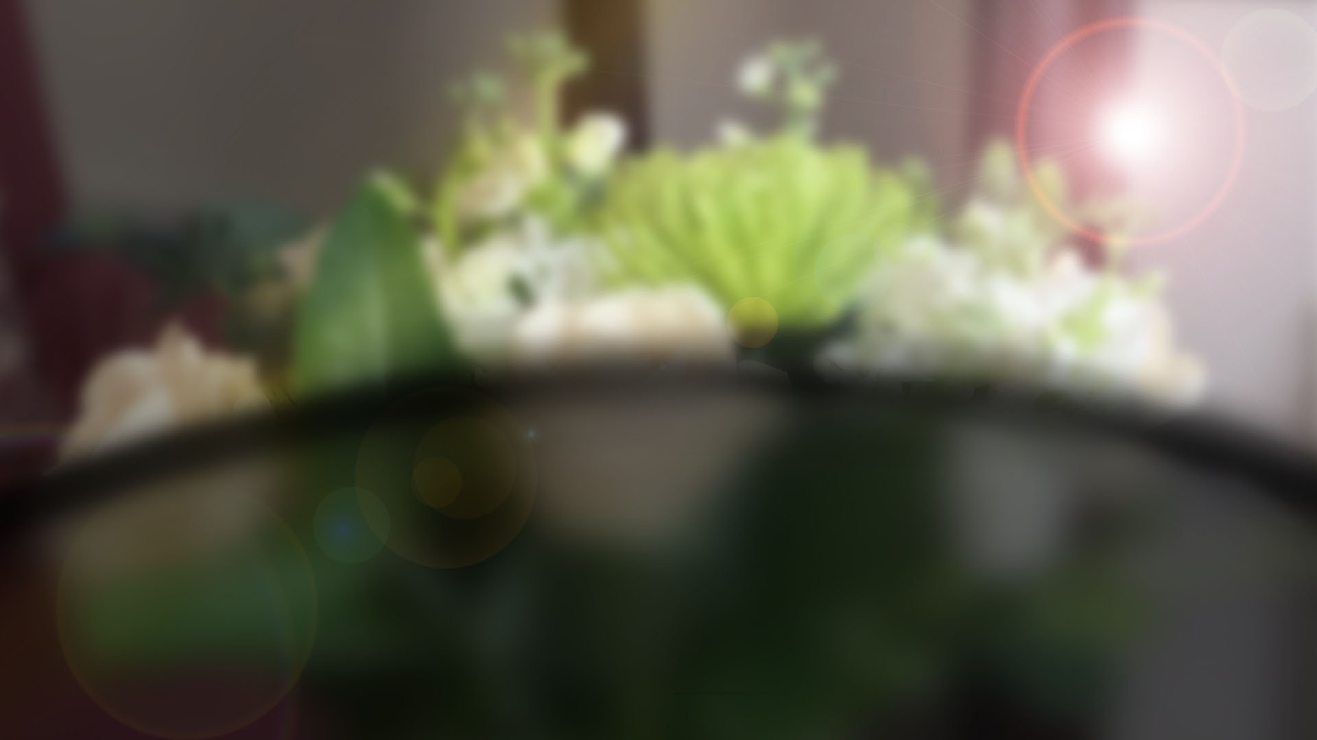
15. Presbyopia 
14. Cataracts 
13. Age-Related Macular Degeneration (AMD) - AdvertisementSwipe or tap ▶ to continue countdown

12. Glaucoma 
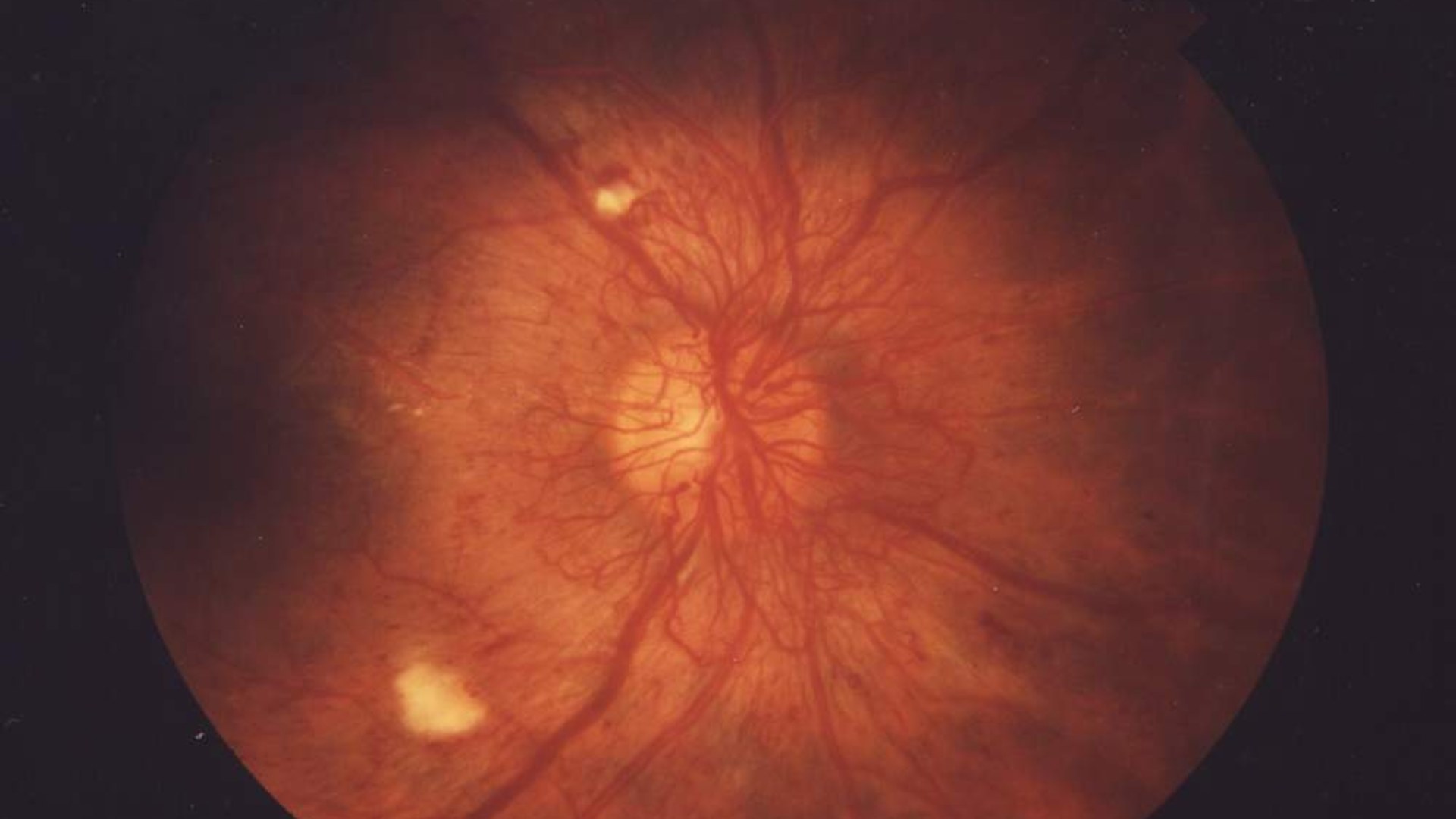
11. Diabetic Retinopathy 
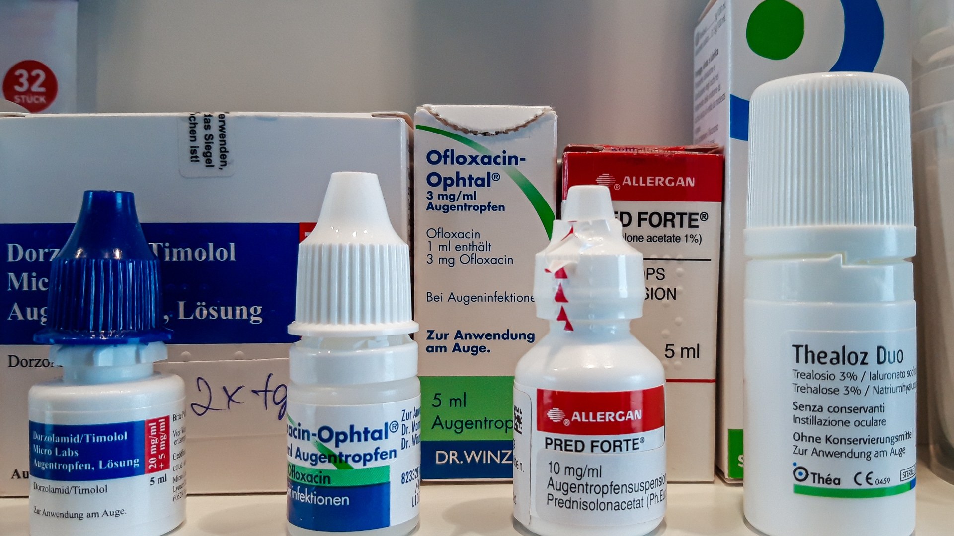
10. Dry Eye Syndrome 
9. Posterior Vitreous Detachment (PVD) - AdvertisementSwipe or tap ▶ to continue countdown

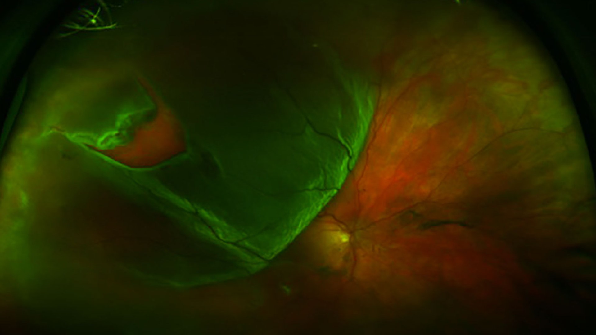
8. Retinal Detachment 
7. Epiretinal Membrane (Macular Pucker) 
6. Retinal Vein Occlusion (Eye Stroke) 
5. Blepharitis - AdvertisementSwipe or tap ▶ to continue countdown

4. Corneal Dystrophies And Degenerations 
3. Uveitis 
2. Ischemic Optic Neuropathy 
1. Ptosis (Drooping Eyelids) - Advertisement🔥 UP NEXT IN THIS SERIES◫ 21 Slides

20 Iconic Historical Photos and the Archive Clues Behind Them
Contact sheets, cracked glass plates, and NASA mission logs preserve the hidden steps behind 20 famous photographs. Drawing on records from the Library of Congress, the National Archives, NASA, and the Smithsonian, this countdown traces how cropping, camera setups, and double exposures shaped each frame.
As the eyes age, structural changes in the lens, cornea, retina, and optic nerve can blur near focus or trigger medical emergencies. From presbyopia and cataracts to silent optic nerve damage and retinal detachment, here are the 15 most common causes of age-related vision loss.
Swipe or tap ▶ to begin the countdown
©Unsplash / Bacila Vlad & Wikimedia / NIH Image Gallery
15. Presbyopia
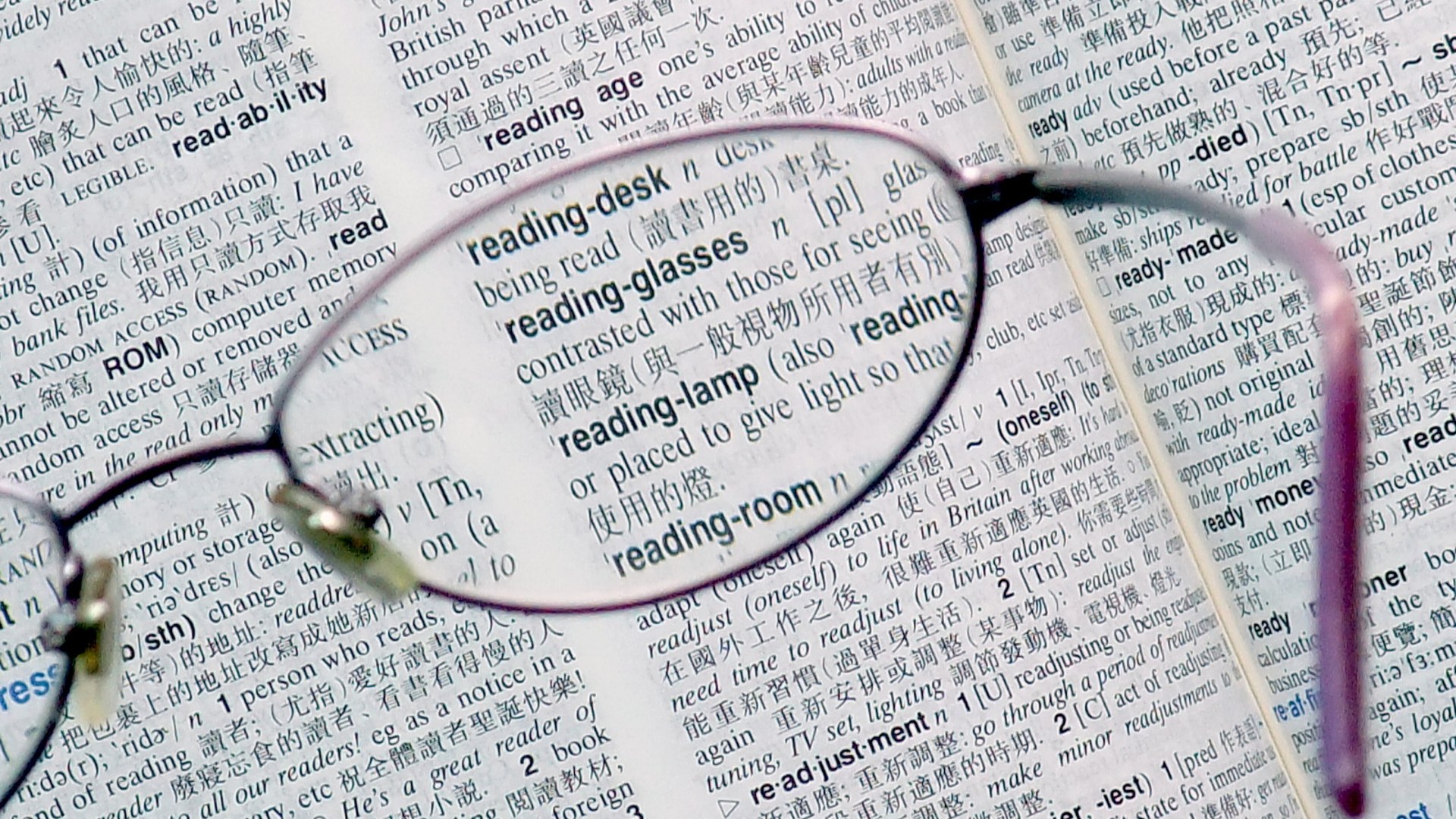
Starting around age 40, the crystalline lens inside the eye gradually loses its flexibility and stiffens, making it harder to focus on close-up text and small print. Reading glasses, bifocals, or multifocal lenses easily restore crisp near vision.
©Wikimedia / Mk2010
14. Cataracts
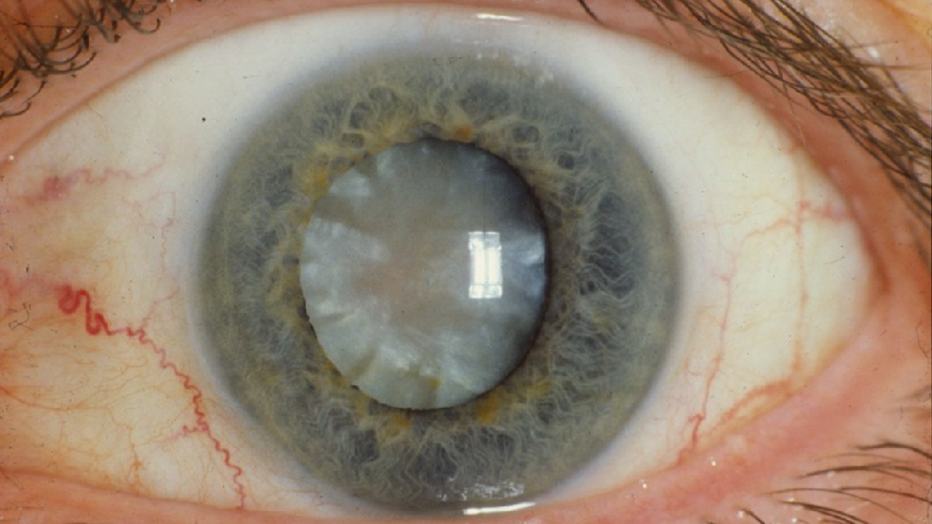
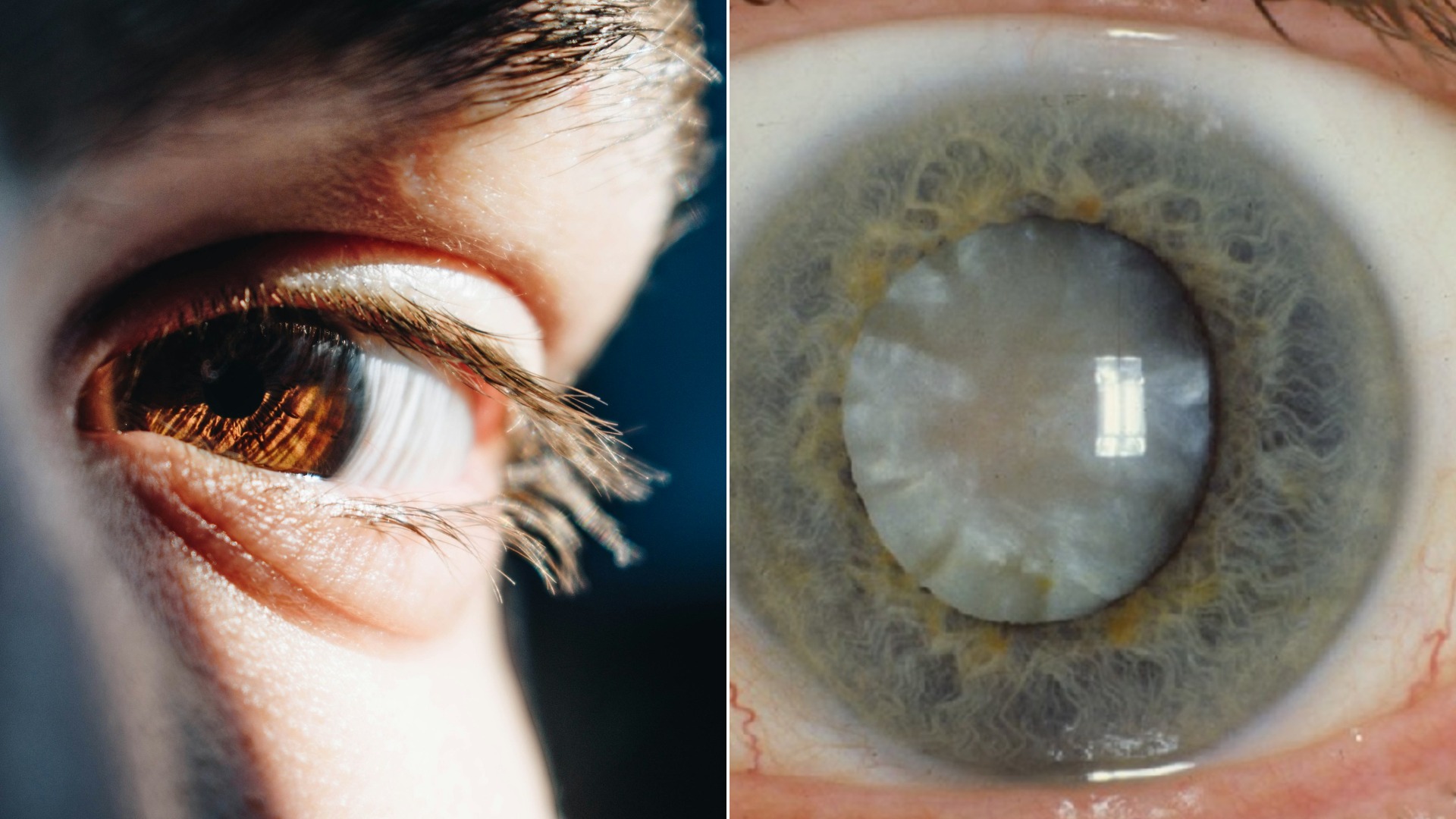
Over decades of UV exposure and oxidative stress, proteins inside the eye's natural lens break down and clump together into cloudy opacities. Colors appear faded, nighttime glare worsens, and sight progressively dims until the clouded lens is replaced surgically.
©Wikimedia / NIH Image Gallery
13. Age-Related Macular Degeneration (AMD)
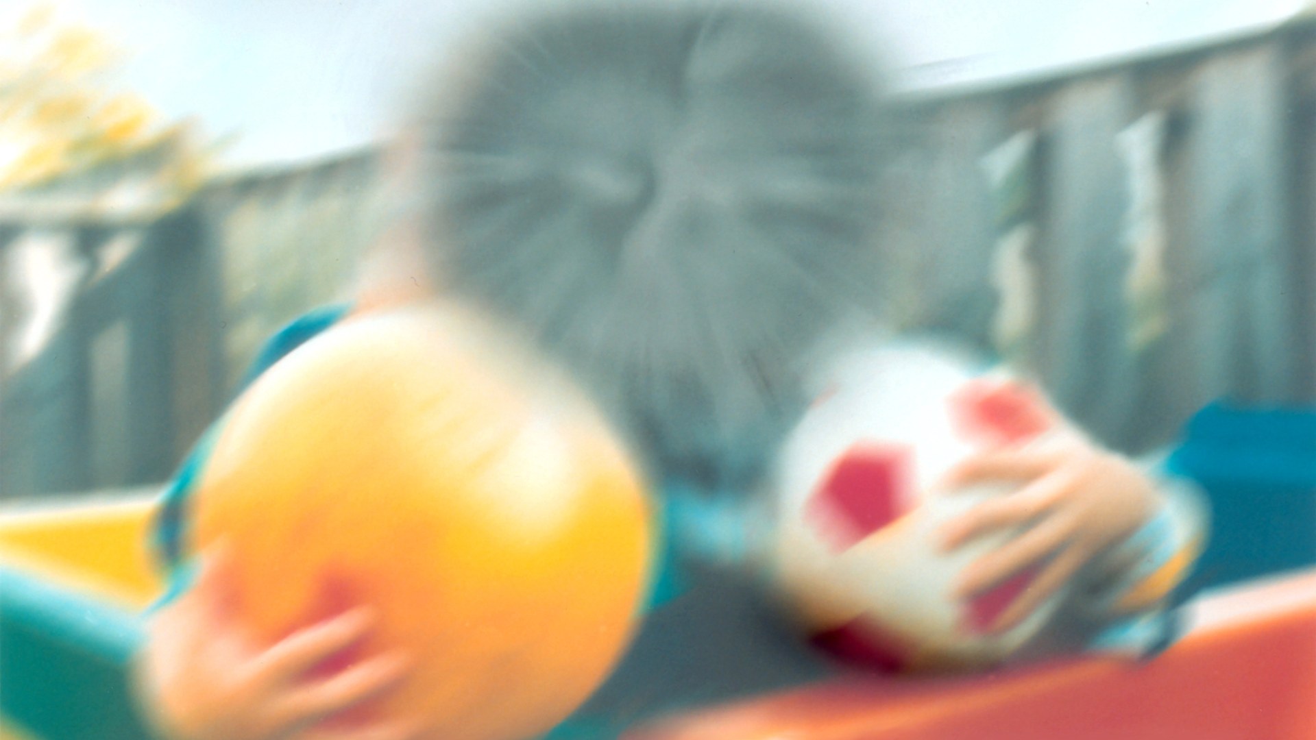
Age-related macular degeneration damages the macula—the tiny central zone of the retina responsible for detailed, straight-ahead sight. While peripheral vision remains intact, dark or blurry blind spots form directly in the center of view, making reading and recognizing faces difficult.
©Wikimedia / National Eye Institute, NIH
Advertisement
Swipe or tap ▶ to resume the countdown.
12. Glaucoma
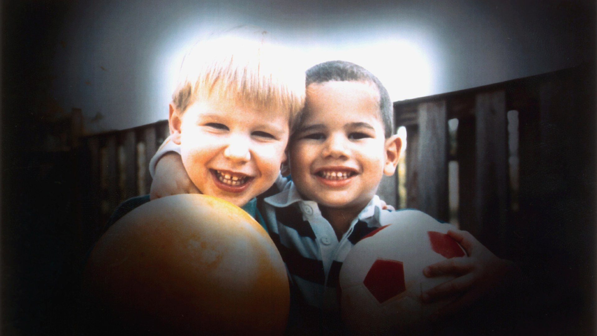
Glaucoma damages the eye's optic nerve—most often when fluid builds up in the front part of the eye and raises intraocular pressure. Because it silently erodes peripheral side vision first without pain or early warning signs, regular eye pressure exams are critical.
©Wikimedia / National Eye Institute, NIH
11. Diabetic Retinopathy
Chronically elevated blood sugar weakens and damages the tiny blood vessels nourishing the light-sensitive retina at the back of the eye. As damaged vessels leak fluid, bleed, or grow fragile scar tissue, vision becomes spotty, blurred, and permanently impaired.
©Wikimedia / National Eye Institute, NIH
10. Dry Eye Syndrome
Tear production naturally declines with age, and changes in the oil glands along the eyelids can cause tears to evaporate too quickly. Without a smooth, stable tear film coating the cornea, eyes feel gritty and inflamed while vision fluctuates throughout the day.
©Wikimedia / Friedrich Haag
9. Posterior Vitreous Detachment (PVD)
As the eye ages, the clear gel-like vitreous humor filling the eyeball gradually liquefies and shrinks until it peels away from the retina. This separation triggers a sudden shower of cobweb-like floaters and brief flashes of light in peripheral vision.
©Wikimedia / Stewart Lamb Cromar
Advertisement
Swipe or tap ▶ to resume the countdown.
8. Retinal Detachment
Retinal detachment occurs when the thin, light-sensitive retinal tissue at the back of the eye tears and pulls away from the nourishing blood vessels beneath it. Often heralded by a dark curtain or shadow sweeping across vision, it requires urgent surgery to prevent permanent blindness.
©Wikimedia / Rocco1963
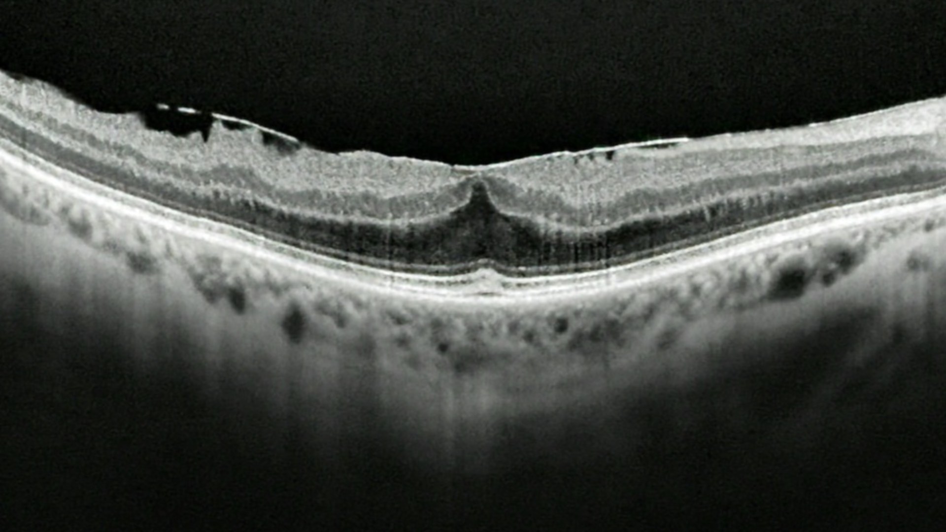
7. Epiretinal Membrane (Macular Pucker)
Often triggered by age-related changes in the vitreous gel, an epiretinal membrane is a delicate sheet of fibrous scar tissue that grows across the surface of the macula. As the membrane contracts, it wrinkles the retina so straight lines appear wavy and fine print looks blurred.
©Wikimedia / Bektelund
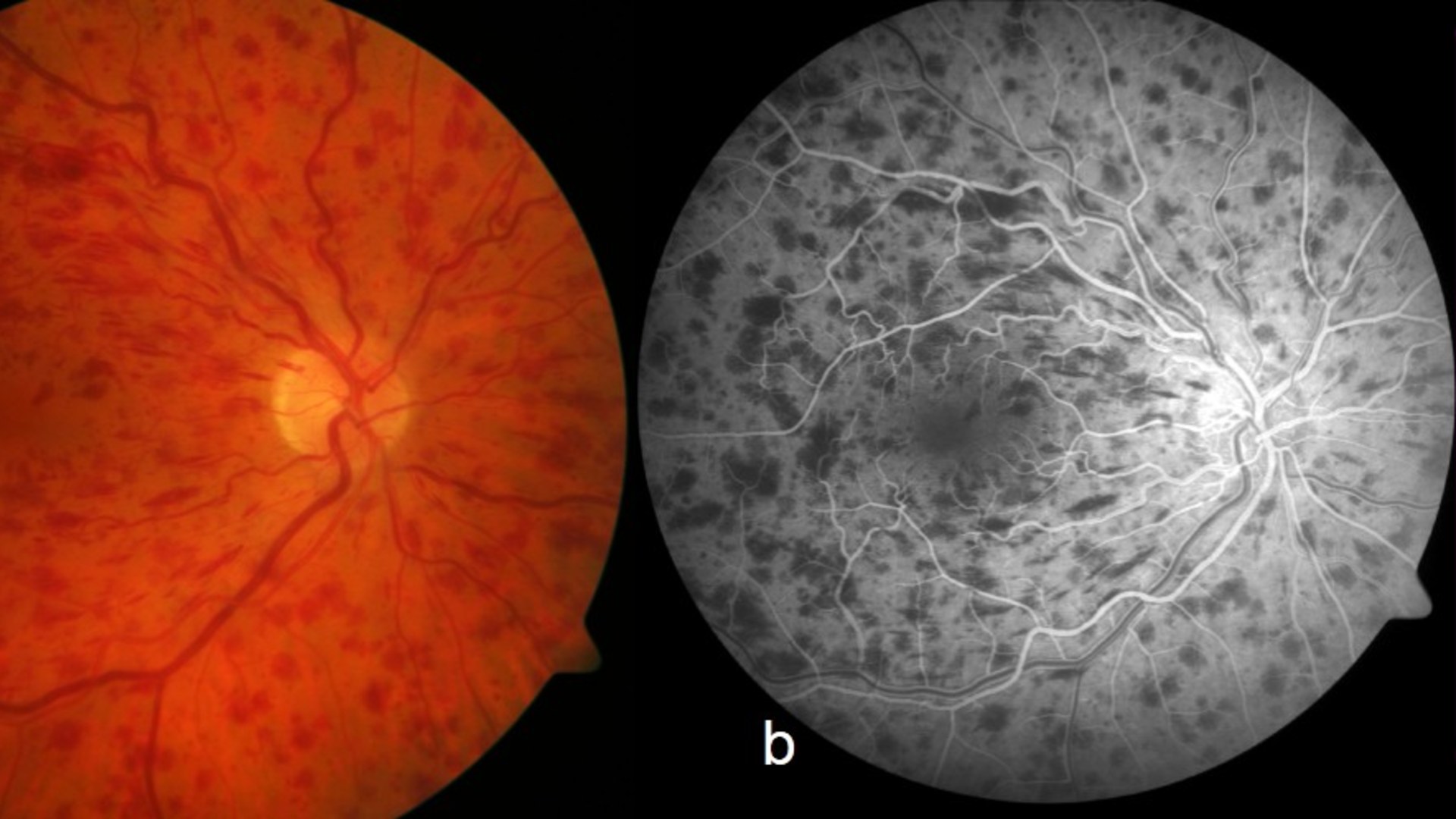
6. Retinal Vein Occlusion (Eye Stroke)
Sometimes called an eye stroke, retinal vein occlusion happens when a blood clot or hardened artery compresses and blocks one of the small veins draining blood from the retina. Blood and fluid back up into the macula, causing sudden, painless blurring or vision loss in one eye.
©Wikimedia / Werner JU et al.
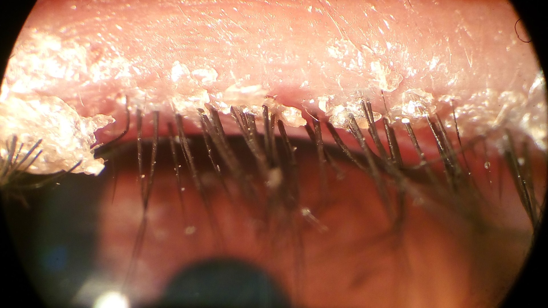
5. Blepharitis
Blepharitis is a stubborn, chronic inflammation along the edges of the eyelids where the eyelashes emerge, often linked to clogged oil glands or skin bacteria. It leaves the eyelids red, crusty, and irritated while destabilizing the tear film and blurring vision.
©Wikimedia / Imrankabirhossain
Advertisement
Swipe or tap ▶ to resume the countdown.
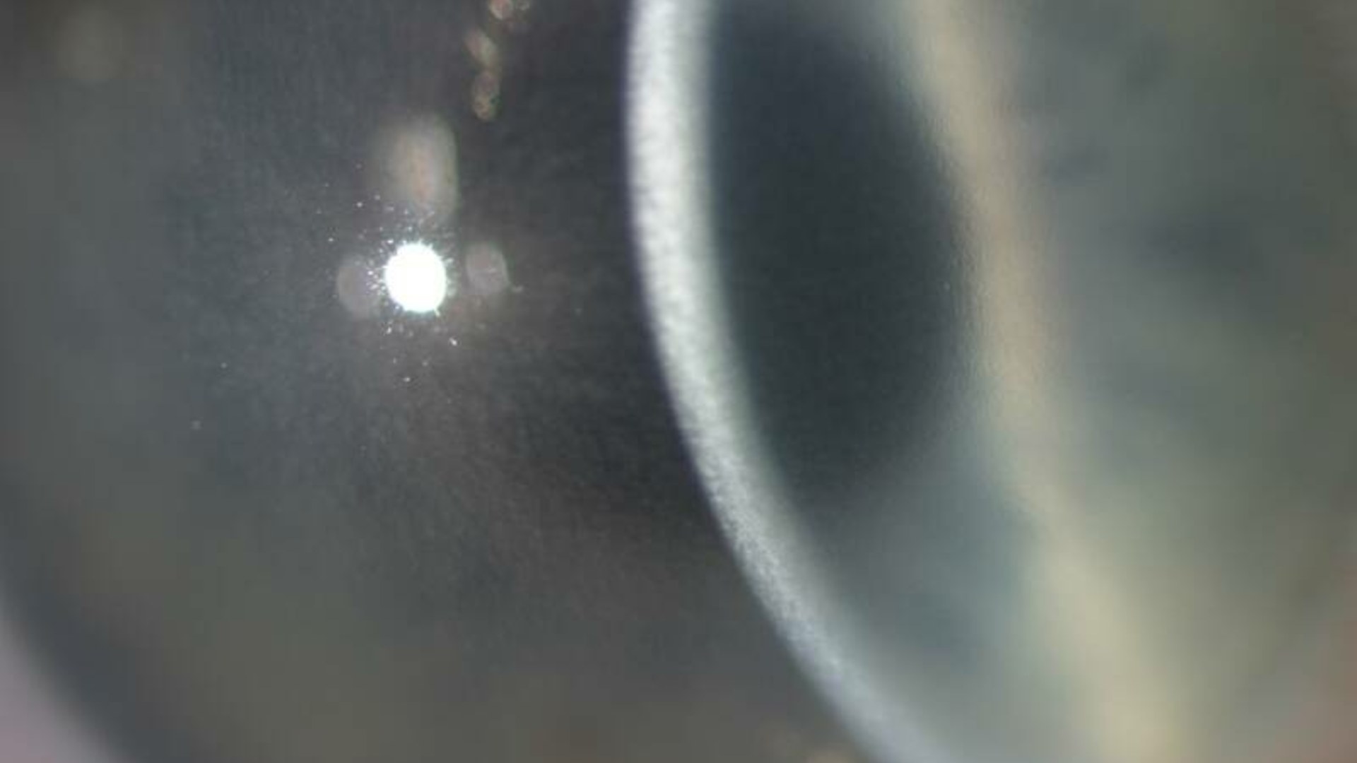
4. Corneal Dystrophies And Degenerations
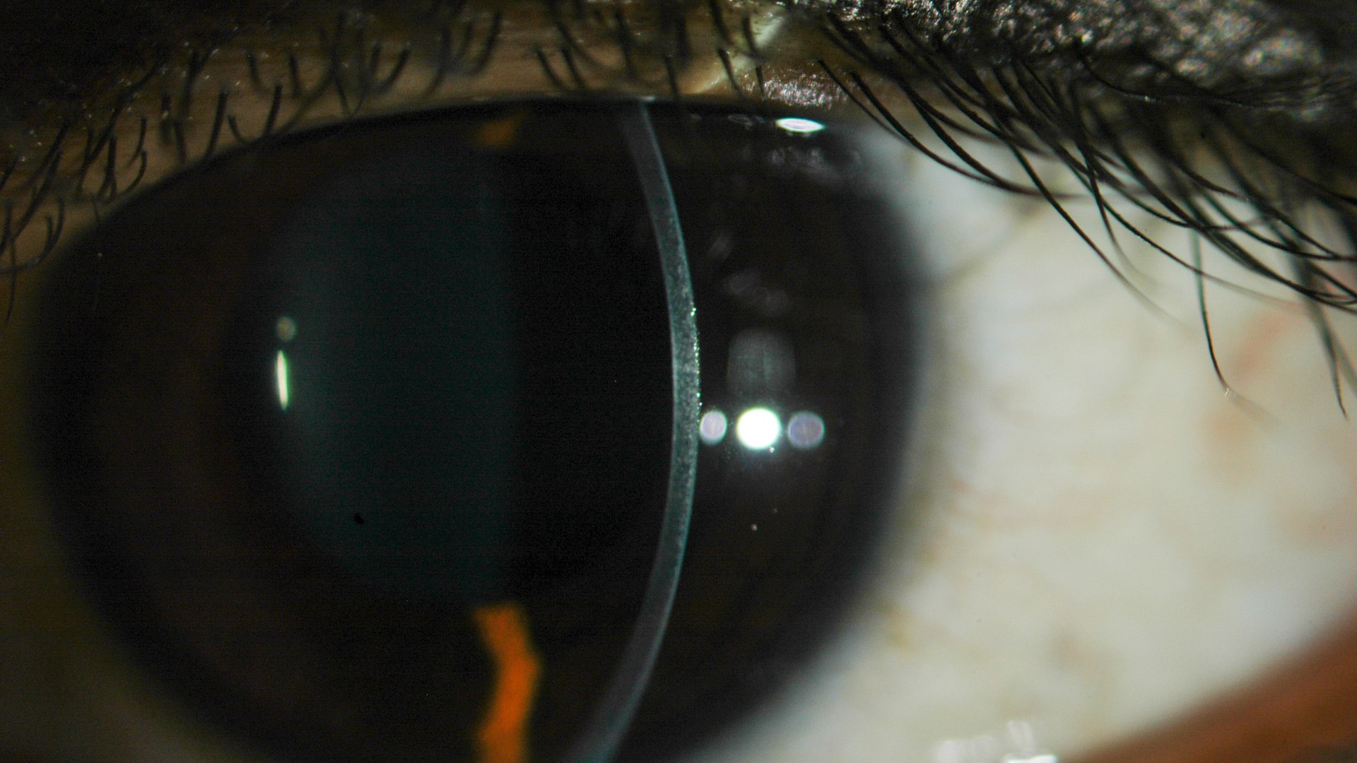
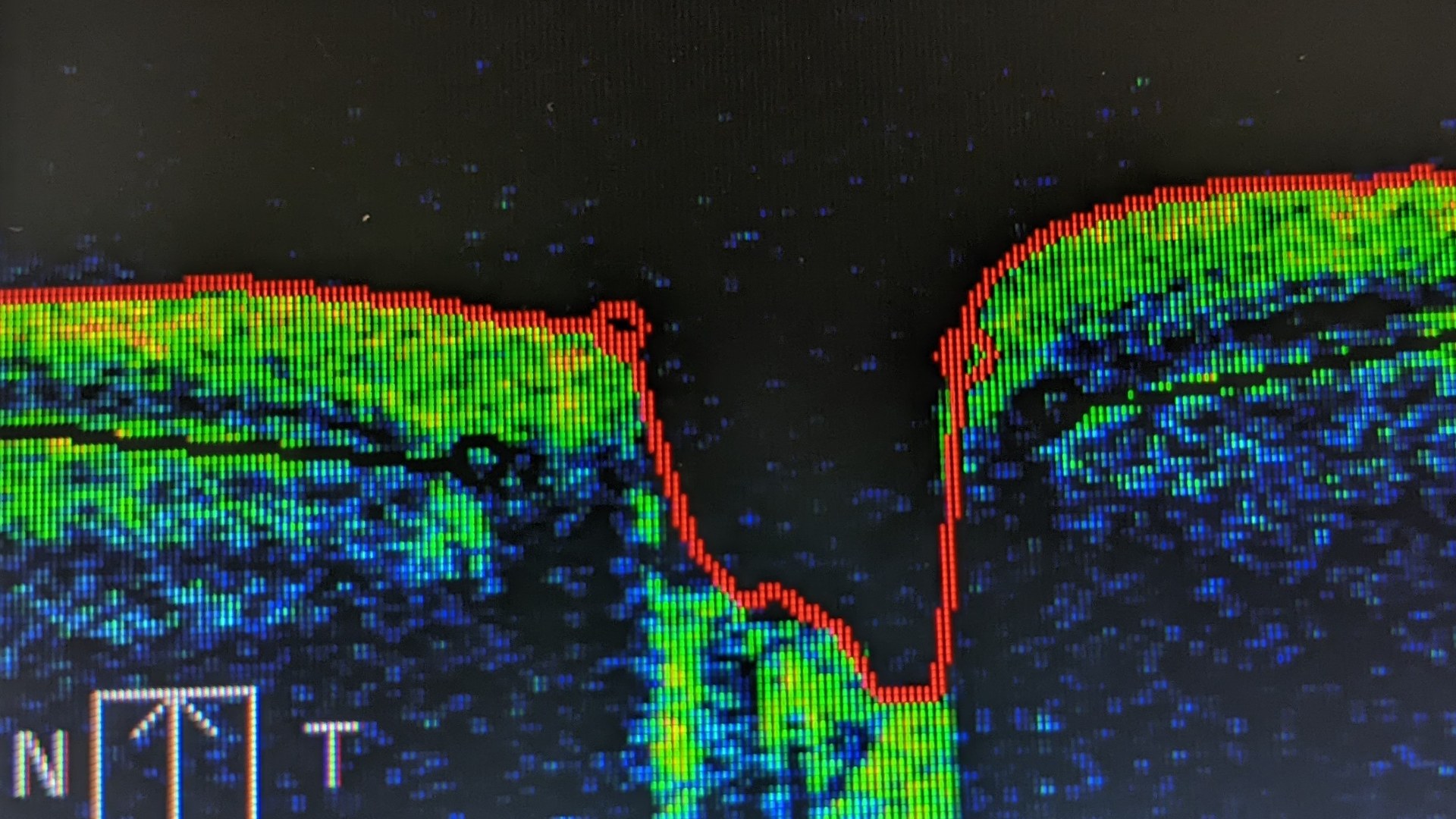
With age, progressive corneal degenerations and dystrophies such as Fuchs' endothelial dystrophy cause fluid or abnormal deposits to accumulate inside the normally crystal-clear outer dome of the eye. As the cornea swells and clouds over, glare increases and visual acuity drops.
©Wikimedia / Klintworth GK.
3. Uveitis
Uveitis is an inflammatory condition affecting the uvea—the pigmented middle layer of the eye wall that supplies blood to the iris, ciliary body, and retina. Left untreated, intraocular inflammation causes eye pain, light sensitivity, floaters, and permanent tissue scarring.
©Wikimedia / Eric Wiessner
2. Ischemic Optic Neuropathy
Ischemic optic neuropathy strikes when blood flow to the optic nerve is suddenly interrupted, starving the nerve fibers that transmit visual signals to the brain. Often associated with hypertension, diabetes, or temporal arteritis in older adults, it causes abrupt, painless vision loss.
©Wikimedia / Bobjgalindo
1. Ptosis (Drooping Eyelids)
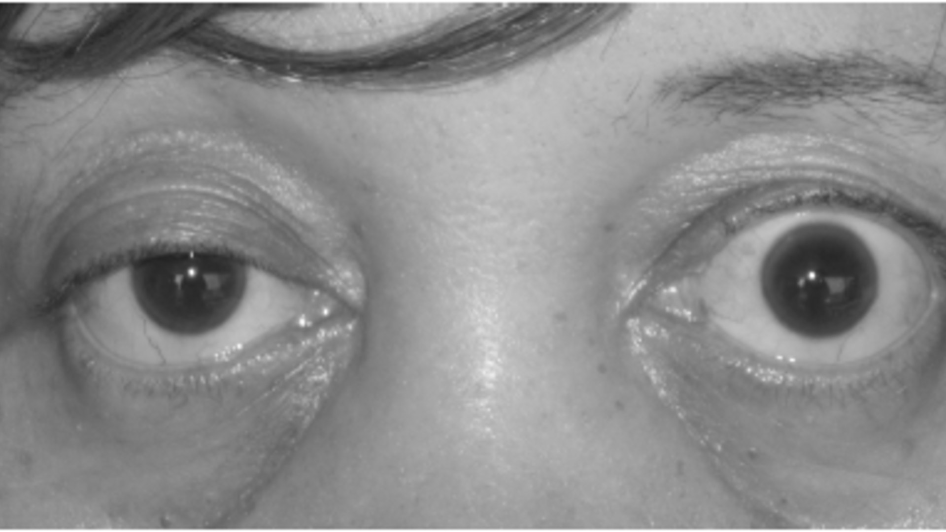
As decades of blinking stretch the levator tendon that lifts the upper eyelid, age-related ptosis causes one or both eyelids to droop lower over the pupil. In severe cases, the sagging lid physically blocks the upper field of vision until surgically elevated.

©Wikimedia / Mohankumar Kurukumbi et al.
Advertisement
You've reached the end of the story. Thanks for reading!

20 Iconic Historical Photos and the Archive Clues Behind Them
Contact sheets, cracked glass plates, and NASA mission logs preserve the hidden steps behind 20 famous photographs. Drawing on records from the Library of Congress, the National Archives, NASA, and the Smithsonian, this countdown traces how cropping, camera setups, and double exposures shaped each frame.
All 15 Entries in this Story
1. Presbyopia
Starting around age 40, the crystalline lens inside the eye gradually loses its flexibility and stiffens, making it harder to focus on close-up text and small print. Reading glasses, bifocals, or multifocal lenses easily restore crisp near vision.

2. Cataracts
Over decades of UV exposure and oxidative stress, proteins inside the eye's natural lens break down and clump together into cloudy opacities. Colors appear faded, nighttime glare worsens, and sight progressively dims until the clouded lens is replaced surgically.

3. Age-Related Macular Degeneration (AMD)
Age-related macular degeneration damages the macula—the tiny central zone of the retina responsible for detailed, straight-ahead sight. While peripheral vision remains intact, dark or blurry blind spots form directly in the center of view, making reading and recognizing faces difficult.

4. Glaucoma
Glaucoma damages the eye's optic nerve—most often when fluid builds up in the front part of the eye and raises intraocular pressure. Because it silently erodes peripheral side vision first without pain or early warning signs, regular eye pressure exams are critical.

5. Diabetic Retinopathy
Chronically elevated blood sugar weakens and damages the tiny blood vessels nourishing the light-sensitive retina at the back of the eye. As damaged vessels leak fluid, bleed, or grow fragile scar tissue, vision becomes spotty, blurred, and permanently impaired.

6. Dry Eye Syndrome
Tear production naturally declines with age, and changes in the oil glands along the eyelids can cause tears to evaporate too quickly. Without a smooth, stable tear film coating the cornea, eyes feel gritty and inflamed while vision fluctuates throughout the day.

7. Posterior Vitreous Detachment (PVD)
As the eye ages, the clear gel-like vitreous humor filling the eyeball gradually liquefies and shrinks until it peels away from the retina. This separation triggers a sudden shower of cobweb-like floaters and brief flashes of light in peripheral vision.

8. Retinal Detachment
Retinal detachment occurs when the thin, light-sensitive retinal tissue at the back of the eye tears and pulls away from the nourishing blood vessels beneath it. Often heralded by a dark curtain or shadow sweeping across vision, it requires urgent surgery to prevent permanent blindness.

9. Epiretinal Membrane (Macular Pucker)
Often triggered by age-related changes in the vitreous gel, an epiretinal membrane is a delicate sheet of fibrous scar tissue that grows across the surface of the macula. As the membrane contracts, it wrinkles the retina so straight lines appear wavy and fine print looks blurred.

10. Retinal Vein Occlusion (Eye Stroke)
Sometimes called an eye stroke, retinal vein occlusion happens when a blood clot or hardened artery compresses and blocks one of the small veins draining blood from the retina. Blood and fluid back up into the macula, causing sudden, painless blurring or vision loss in one eye.

11. Blepharitis
Blepharitis is a stubborn, chronic inflammation along the edges of the eyelids where the eyelashes emerge, often linked to clogged oil glands or skin bacteria. It leaves the eyelids red, crusty, and irritated while destabilizing the tear film and blurring vision.

12. Corneal Dystrophies And Degenerations
With age, progressive corneal degenerations and dystrophies such as Fuchs' endothelial dystrophy cause fluid or abnormal deposits to accumulate inside the normally crystal-clear outer dome of the eye. As the cornea swells and clouds over, glare increases and visual acuity drops.

13. Uveitis
Uveitis is an inflammatory condition affecting the uvea—the pigmented middle layer of the eye wall that supplies blood to the iris, ciliary body, and retina. Left untreated, intraocular inflammation causes eye pain, light sensitivity, floaters, and permanent tissue scarring.

14. Ischemic Optic Neuropathy
Ischemic optic neuropathy strikes when blood flow to the optic nerve is suddenly interrupted, starving the nerve fibers that transmit visual signals to the brain. Often associated with hypertension, diabetes, or temporal arteritis in older adults, it causes abrupt, painless vision loss.

15. Ptosis (Drooping Eyelids)
As decades of blinking stretch the levator tendon that lifts the upper eyelid, age-related ptosis causes one or both eyelids to droop lower over the pupil. In severe cases, the sagging lid physically blocks the upper field of vision until surgically elevated.